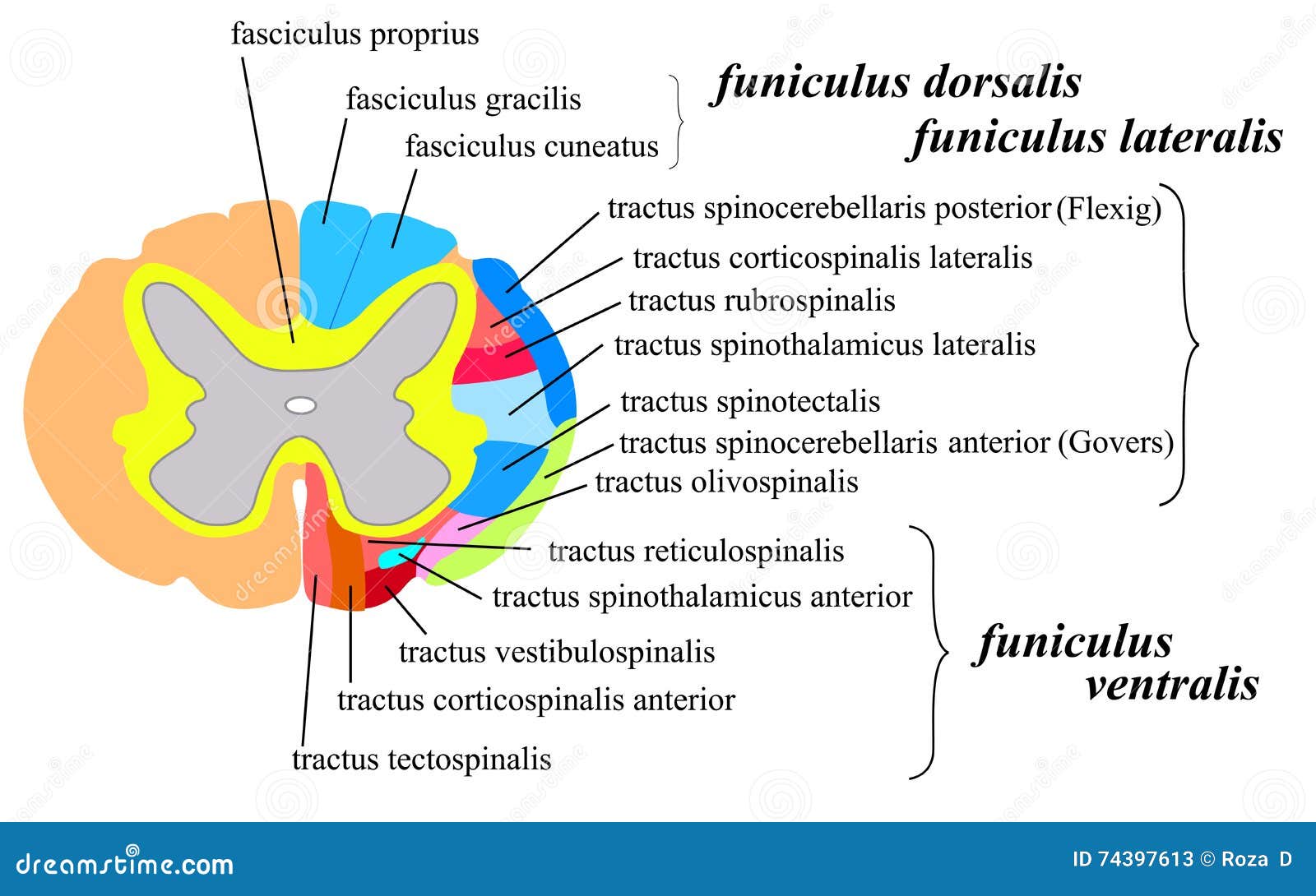
peripheral nerve cross section
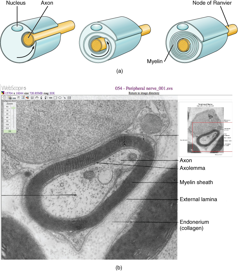
This three-part diagram shows the process of myelination. In step A. 9 Pictures about This three-part diagram shows the process of myelination. In step A : Nervous System - Slide #14, Nervous Tissue and also This three-part diagram shows the process of myelination. In step A.
This Three-part Diagram Shows The Process Of Myelination. In Step A
 oerpub.github.io
myelin sheath myelination tissue process neurilemma myelinated nerve nervous difference between cell axon anatomy figure fibers around schwann cells neuron
Nervous Tissue
 histologydrawings.blogspot.com
nervous system nerve peripheral histology hematoxylin eosin tissue anatomy human physiology slides medical drawings spinal spinalis medulla neurons cord students
Nervous System: The Spinal Cord White Matter Cartoon Vector
 cartoondealer.com
spinal cord matter section cross conductive side nervous system cords path rear clipart medical
Distribution Of A Spinal Nerves | ClipArt ETC
 etc.usf.edu
spinal nerves distribution etc clipart medium usf edu
Cross Section Of The Spinal Cord Royalty Free Stock Photos - Image
 www.dreamstime.com
spinale midollo trasversale peripheral dwarsdoorsnede ruggemerg cervical lumbar myelopathy nerves neurons sympathetic spondylotic pns rückenmarks querschnitt nerf pelviche nervose radici
Tkanka Nerwowa I Tkanka Glejowa
 eszkola.pl
tkanka nerwowa reytan mikroskopowy
Nerves: The Histology Guide
 histology.leeds.ac.uk
histology nerves nerve peripheral perineurium pns section endoneurium opposite stained labels pink help
Nervous System - Slide #14
 education.med.nyu.edu
section cross nerve histology nervous bundle trichrome mallory system slide epineurium endoneurium myelin nyu med education edu 25x sheath slides
1 General Principles Of Ultrasound-Guided Peripheral Nerve Blocks
 aneskey.com
nerve structure peripheral guided principles ultrasound blocks general fig et al source
Nervous system nerve peripheral histology hematoxylin eosin tissue anatomy human physiology slides medical drawings spinal spinalis medulla neurons cord students. This three-part diagram shows the process of myelination. in step a. 1 general principles of ultrasound-guided peripheral nerve blocks