pelvis hip anatomy
Developmental dysplasia of the hip | Image | Radiopaedia.org. 9 Pictures about Developmental dysplasia of the hip | Image | Radiopaedia.org : Interpreting X-Rays of the Pelvis, Hip Joint and Femur - YouTube, 👨🏽💻Want to learn a system for reviewing a pelvic X-ray? Read on to and also Interpreting X-Rays of the Pelvis, Hip Joint and Femur - YouTube.
Developmental Dysplasia Of The Hip | Image | Radiopaedia.org
 radiopaedia.org
hip dysplasia developmental radiopaedia
Vascular Anatomy Of Groin Medical Illustration Medivisuals | Vascular
 www.pinterest.com
anatomy groin neurovascular vascular femoral arteries pelvis illustration veins nerve artery vein medivisuals1 medical system circulatory saphenous superficial
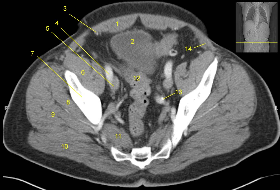
Pelvis Computed Tomograph (axial CT)
 jhurads4anatomy.com
ct pelvis anatomy scan pelvic muscle axial iliacus bone labeled computed cat tomograph legend study ss1 docs storage google
Small Intestine: Anatomy, Location And Function | Kenhub
intestine anatomy location function kenhub background
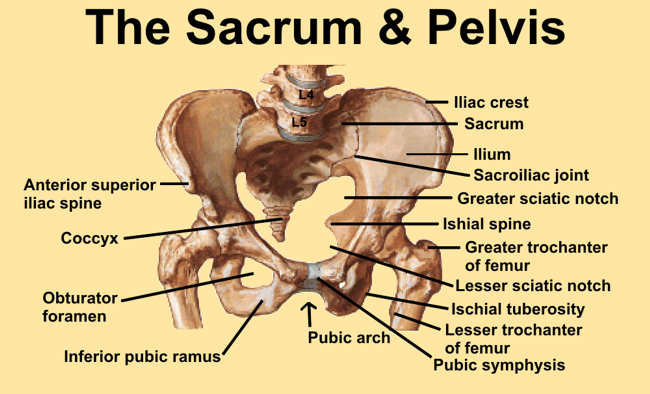
Pelvic Fractures - Physiopedia, Universal Access To Physiotherapy
 www.physio-pedia.com
pelvis diagram pelvic anatomy sacrum bones landmarks hip human bone bony girdle fractures skeletal skeleton physio pedia joint series identified
Hip Case 2 - Sports Medicine Imaging
 sportsmedicineimaging.com
hip labral case sports spine imaging
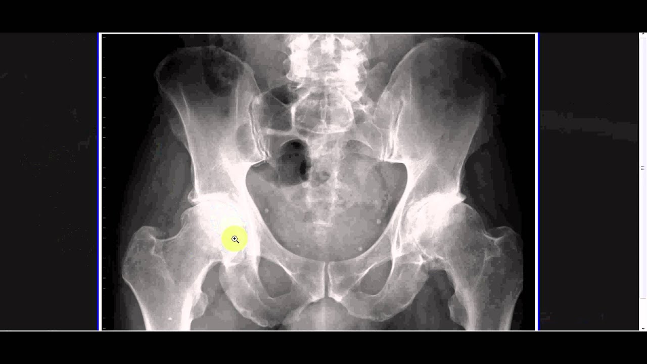
👨🏽💻Want To Learn A System For Reviewing A Pelvic X-ray? Read On To
 www.pinterest.com
ray pelvic anatomy radiology left
Interpreting X-Rays Of The Pelvis, Hip Joint And Femur - YouTube
 www.youtube.com
hip xray female joint pain pelvis femur interpreting rays anatomy pelvic arthritis bone
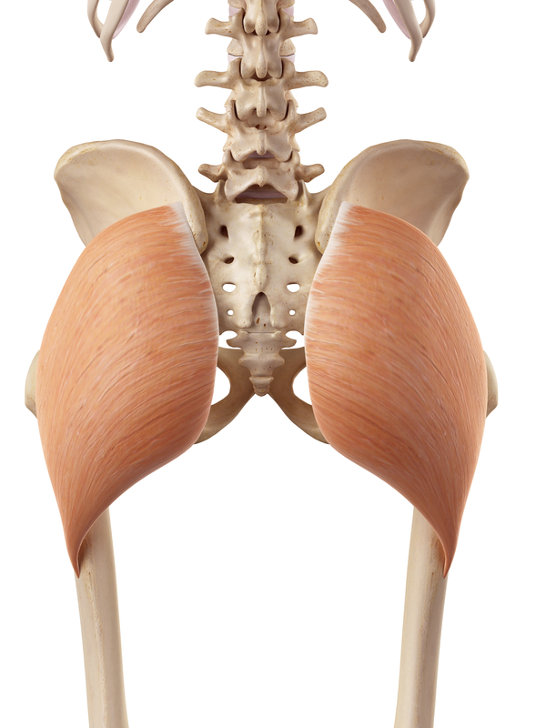
The Gluteus Maximus And Hip Extension
 healthybackandwellnessinstitute.com
gluteus maximus hip illustration glute max extension muscle medical
Developmental dysplasia of the hip. Hip xray female joint pain pelvis femur interpreting rays anatomy pelvic arthritis bone. Vascular anatomy of groin medical illustration medivisuals