orbit bone anatomy
Ethmoid bone: Anatomy, borders and development | Kenhub. 9 Pictures about Ethmoid bone: Anatomy, borders and development | Kenhub : Magnified Anterior View of Orbital Apex | Neuroanatomy | The, Anatomy For Sculptors - The orbital region forms by anatomy for sculptors and also Brachial artery: Anatomy and branches | Kenhub.
Ethmoid Bone: Anatomy, Borders And Development | Kenhub
bone ethmoid concha anatomy kenhub nasal inferior os ethmoidale skull nodes nasalis pelvis lymphatic bones head vessels lateral cavity structure
Frontal Bone: Anatomy, Borders And Development | Kenhub
frontal bone kenhub anatomy frontale os
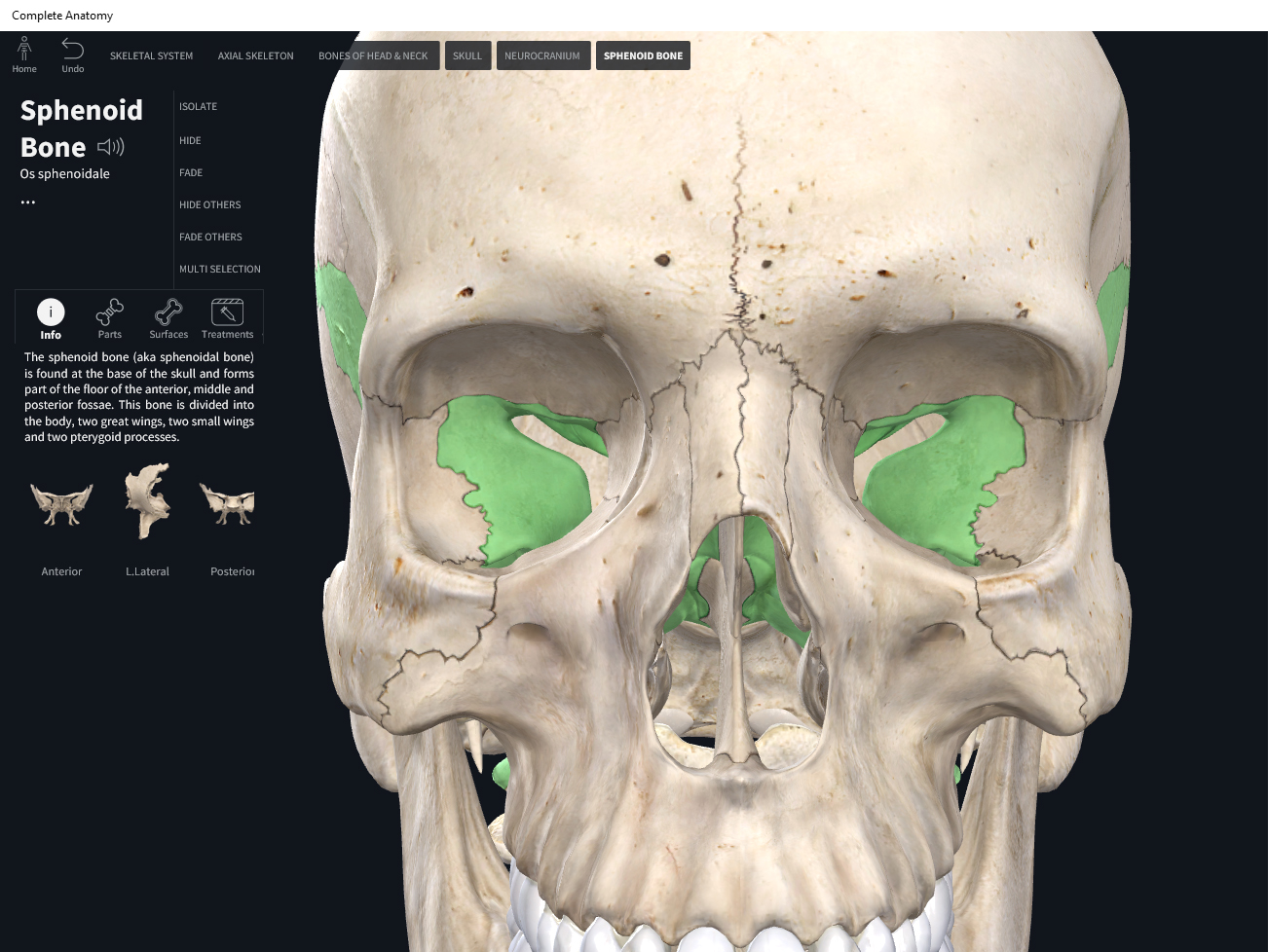
Bones: Skull, Sphenoid. – Anatomy & Physiology
 integrativewellnessandmovement.com
sphenoid references
Brachial Artery: Anatomy And Branches | Kenhub
artery brachial axillary anatomy subclavian muscle kenhub vein nerve arteria branches arm region subclavia superior ulnar brachialis axillaris arteries collateral
Eosinophilic Granuloma — Clinical MRI
 clinical-mri.com
granuloma eosinophilic mri clinical
Metastasis (carcinoma Of Prostate) — Clinical MRI
 clinical-mri.com
metastasis prostate mri carcinoma t1 clinical t2
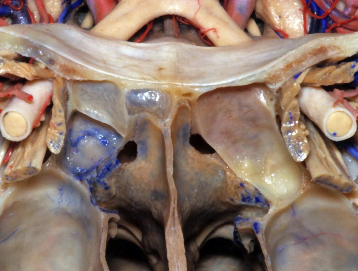
Magnified Anterior View Of Orbital Apex | Neuroanatomy | The
 www.neurosurgicalatlas.com
orbital apex magnified neurosurgicalatlas surgical
Anatomy For Sculptors - The Orbital Region Forms By Anatomy For Sculptors
 anatomy4sculptors.artstation.com
anatomy sculptors region orbital eye artstation neck forms orbit sculpture face anatomy4sculptors human artists form rthe drawing head eyelids shapes
Enchondroma — Clinical MRI
 clinical-mri.com
enchondroma mri clinical
Bones: skull, sphenoid. – anatomy & physiology. Enchondroma — clinical mri. Brachial artery: anatomy and branches