occipital bone veterinary anatomy
Oral Anatomy of the Dog and Cat in Veterinary Dentistry Practice. 9 Images about Oral Anatomy of the Dog and Cat in Veterinary Dentistry Practice : Posterior Neck and Occipital Region | Plastic Surgery Key, Chiari-Like Malformation (CM) & Syringomyelia (SM) and the Cavalier and also Anatomy and Physiology Nursing Mnemonics & Tips.
Oral Anatomy Of The Dog And Cat In Veterinary Dentistry Practice
 www.vetsmall.theclinics.com
cranium ventral
Canine Radiographs
 www.slideshare.net
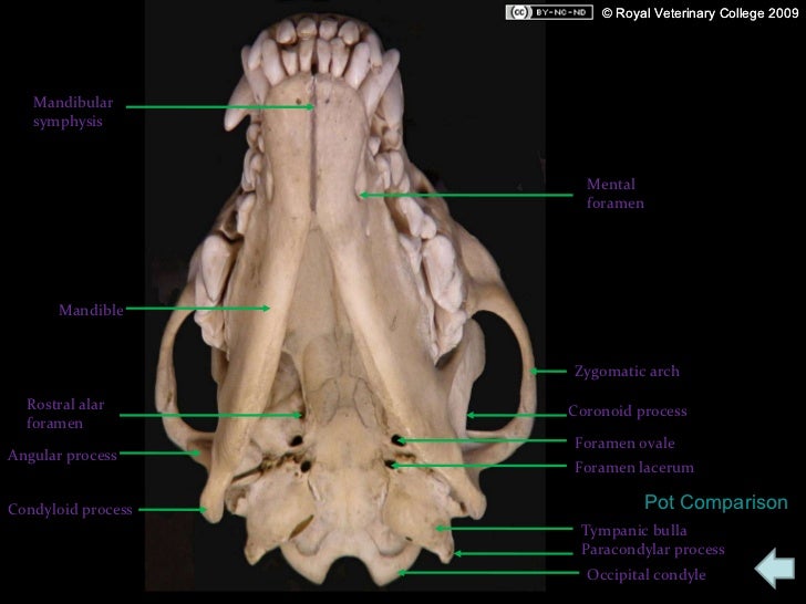
canine radiographs foramen mandibular symphysis mandible alar condyloid rostral angular
Anatomy Of The Horse: Osteology
 www.imaios.com
horse anatomy ribs thoracic skeleton equine sternum costal cartilage osteology skeletal system atlas animal bones vet horses imaios visit
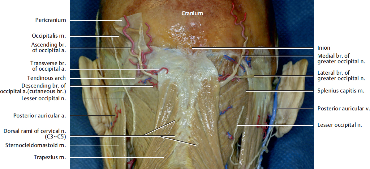
Posterior Neck And Occipital Region | Plastic Surgery Key
 plasticsurgerykey.com
occipital posterior region neck muscle left galea skin side occipitalis surgery fig been plasticsurgerykey
Occipital Nerve Blocks | Anesthesia Key
 aneskey.com
nerve occipital blocks block
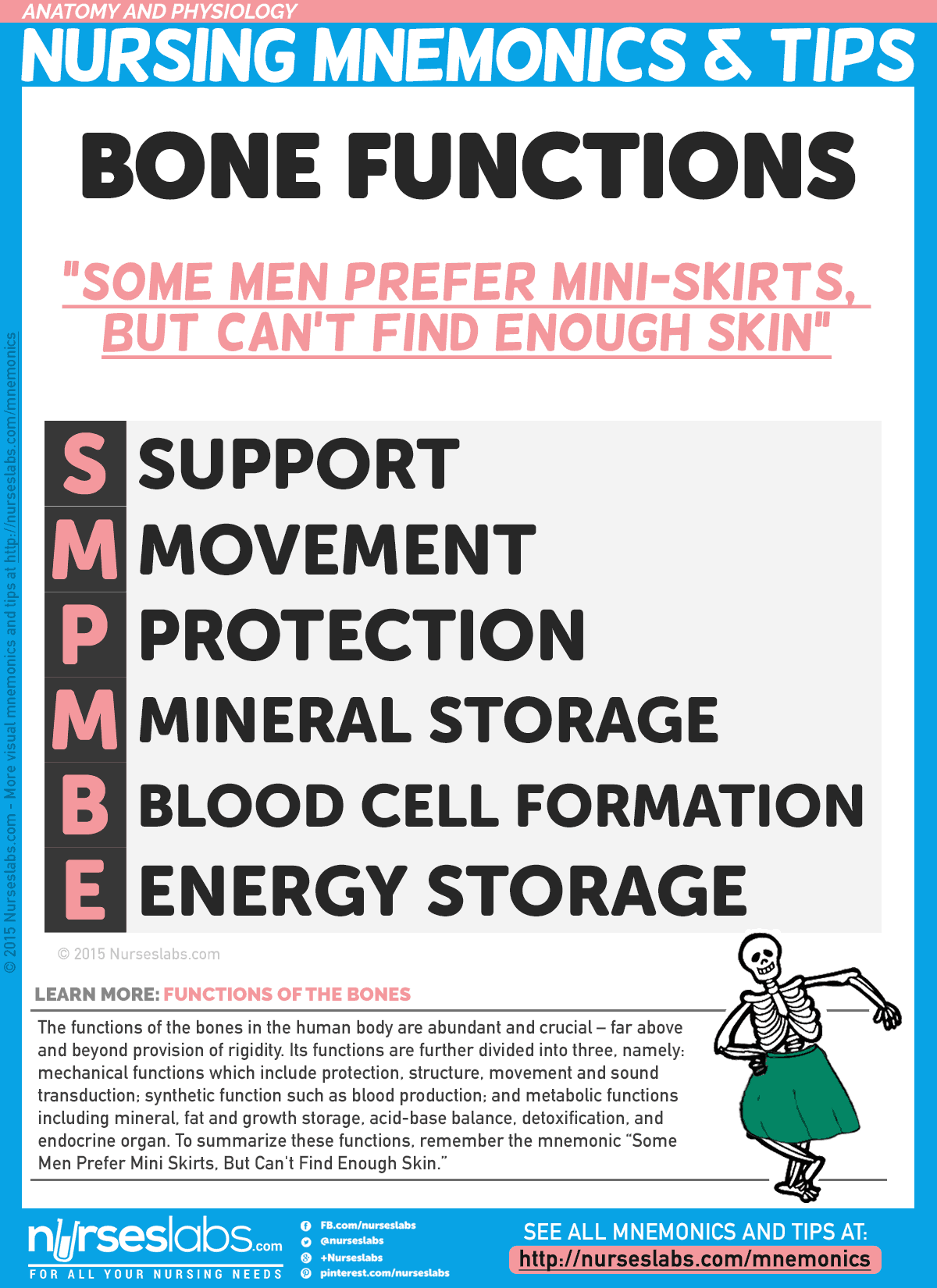
Anatomy And Physiology Nursing Mnemonics & Tips
 nurseslabs.com
functions bone mnemonics anatomy bones nursing physiology nurseslabs skin tips mnemonic remember mm
Image Gallery: 3D Rat Anatomy Software - Biosphera
 biosphera3d.com
masseter testis uterus spinal
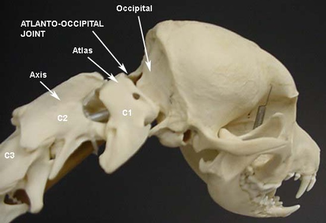
Chiari-Like Malformation (CM) & Syringomyelia (SM) And The Cavalier
 cavalierhealth.com
charles king atlas occipital skull cavalier spaniel bone atlanto joint foramen magnum vertebra syringomyelia c1 dog sm neck malformation ligaments
Temporal Bone Surgery | Neupsy Key
 neupsykey.com
bone
Anatomy of the horse: osteology. Canine radiographs foramen mandibular symphysis mandible alar condyloid rostral angular. Image gallery: 3d rat anatomy software