nerves in foot diagram
anatomical landmarks on the dorsum of the left foot showing surface. 9 Pictures about anatomical landmarks on the dorsum of the left foot showing surface : anatomical landmarks on the dorsum of the left foot showing surface, Blood Supply to the Leg and Foot – Human Anatomy for Physician and also Cardiac muscle tissue histology | Kenhub.
Anatomical Landmarks On The Dorsum Of The Left Foot Showing Surface
dorsum anatomical edb
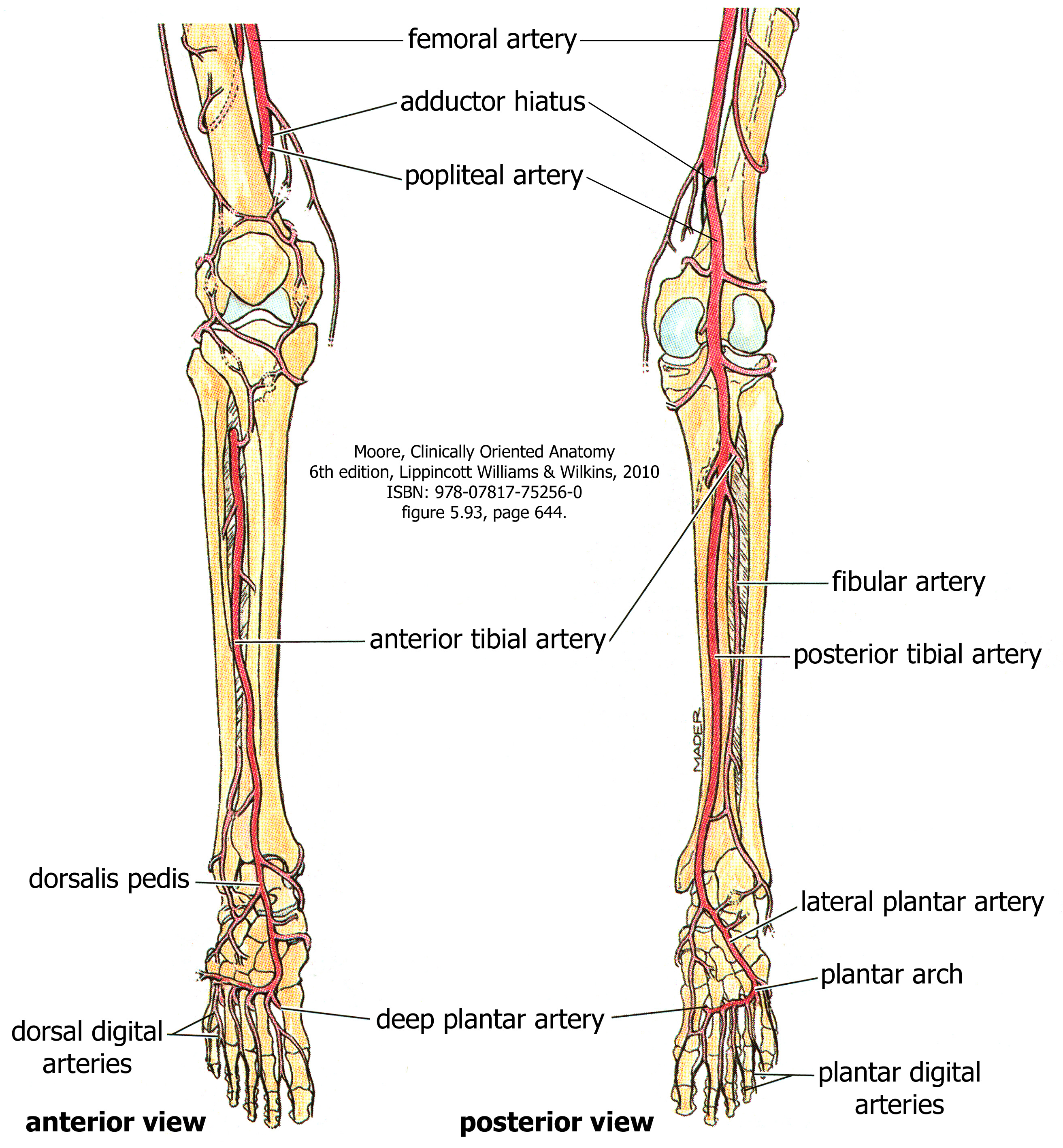
Blood Supply To The Leg And Foot – Human Anatomy For Physician
 wisc.pb.unizin.org
arteries collateral limbs
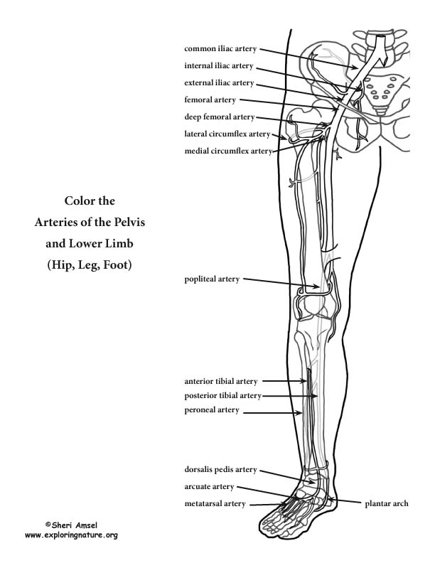
Arteries Of The Lower Limb (Pelvis, Leg And Foot) Coloring Page
 www.exploringnature.org
lower coloring arteries limb leg anatomy foot pelvis pdf hip human diagram template upper physiology muscle blank labeling advanced exploringnature
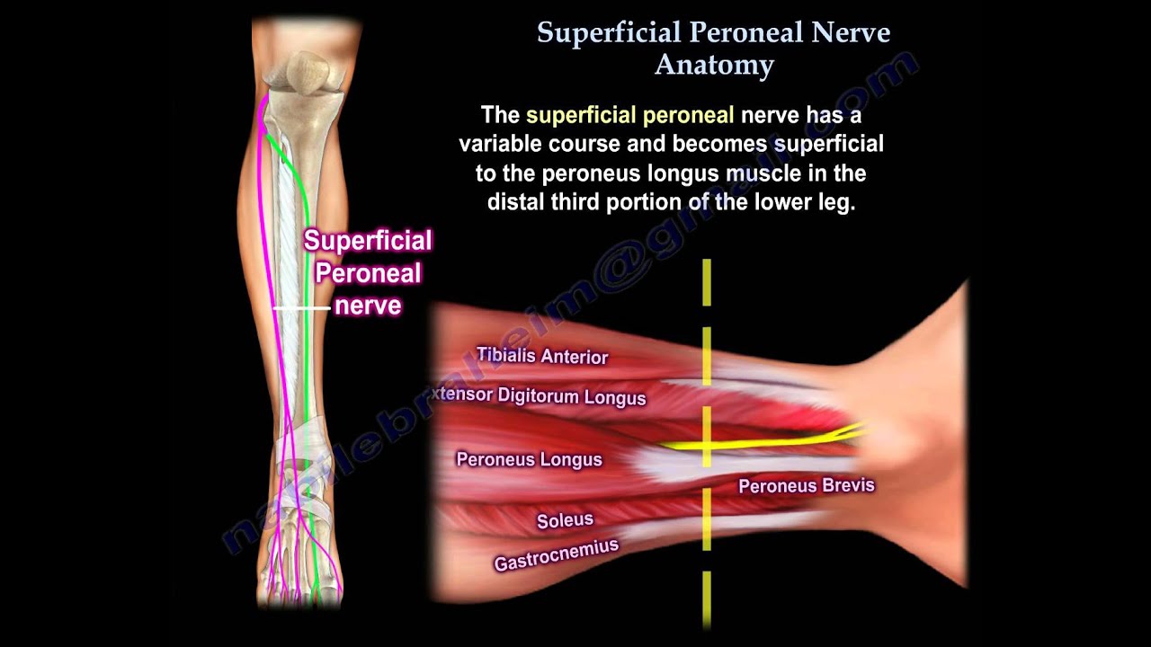
Superficial Peroneal Nerve Anatomy - Everything You Need To Know - Dr
 www.youtube.com
nerve peroneal superficial anatomy
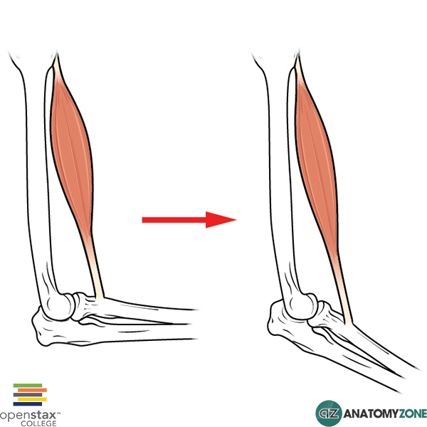
Eccentric Muscle Contraction • Muscular, Musculoskeletal • AnatomyZone
 anatomyzone.com
contraction eccentric concentric anatomyzone isometric
Foot Anatomy | EOrthopod.com
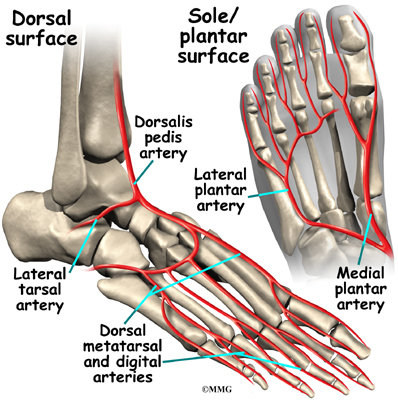 www.eorthopod.com
pedis dorsalis tibialis artery arteries vessels works eorthopod bohn
Ankle Anatomy | EOrthopod.com
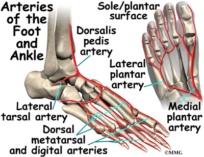 eorthopod.com
ankle anatomy foot toe arteries brachial eorthopod legs blood supply artery diagram joint feet lateral flow inside anterior syndrome muscles
Shoulder Joint Cross Section - Medical Art Library
 medicalartlibrary.com
joint shoulder anatomy glenohumeral section cross glenoid cavity labrum humeral head diagram medical fossa medicalartlibrary joints illustration select category
Cardiac Muscle Tissue Histology | Kenhub
cardiac tissue muscle histology heart slide histological kenhub striated muscles structure anatomy cells myocardium types tissues fiber function location library
Contraction eccentric concentric anatomyzone isometric. Cardiac tissue muscle histology heart slide histological kenhub striated muscles structure anatomy cells myocardium types tissues fiber function location library. Anatomical landmarks on the dorsum of the left foot showing surface